Beranda
/ Upper Thigh Anatomy / Upper Legs Running Anatomy Sports Anatomy / If talking about the skull, the dorsal side is the top.
Upper Thigh Anatomy / Upper Legs Running Anatomy Sports Anatomy / If talking about the skull, the dorsal side is the top.
Insurance Gas/Electricity Loans Mortgage Attorney Lawyer Donate Conference Call Degree Credit Treatment Software Classes Recovery Trading Rehab Hosting Transfer Cord Blood Claim compensation mesothelioma mesothelioma attorney Houston car accident lawyer moreno valley can you sue a doctor for wrong diagnosis doctorate in security top online doctoral programs in business educational leadership doctoral programs online car accident doctor atlanta car accident doctor atlanta accident attorney rancho Cucamonga truck accident attorney san Antonio ONLINE BUSINESS DEGREE PROGRAMS ACCREDITED online accredited psychology degree masters degree in human resources online public administration masters degree online bitcoin merchant account bitcoin merchant services compare car insurance auto insurance troy mi seo explanation digital marketing degree floridaseo company fitness showrooms stamfordct how to work more efficiently seowordpress tips meaning of seo what is an seo what does an seo do what seo stands for best seotips google seo advice seo steps, The secure cloud-based platform for smart service delivery. Safelink is used by legal, professional and financial services to protect sensitive information, accelerate business processes and increase productivity. Use Safelink to collaborate securely with clients, colleagues and external parties. Safelink has a menu of workspace types with advanced features for dispute resolution, running deals and customised client portal creation. All data is encrypted (at rest and in transit and you retain your own encryption keys. Our titan security framework ensures your data is secure and you even have the option to choose your own data location from Channel Islands, London (UK), Dublin (EU), Australia.
Upper Thigh Anatomy / Upper Legs Running Anatomy Sports Anatomy / If talking about the skull, the dorsal side is the top.. Medial muscles adduct and rotate your thigh, and posterior flex your leg and extend your thigh. They work closely with your quadriceps muscles at the front of your thigh, your gluteal muscles, and your calf muscles to ensure proper movement of your leg and hip. Nestled into the top part of the pelvic bone, the iliacus muscle extends all the way from your lowest rib to your thigh bone. Legs give us the freedom to run, walk, jump, climb, and negotiate the world around us. Related links to external sites (from bing) these images are a random sampling from a bing search on the term vascular anatomy of the thigh.
See the pictures and anatomy description of knee joint bones, cartilage, ligaments, muscle and tendons with resources for knee problems & injuries. The hamstring portion of the adductor magnus has a similar action to these muscles, but is located in the medial thigh. Check out the diagram of leg muscle anatomy in the first diagram below. Related posts of muscle anatomy of upper thigh. Похожие запросы для upper thigh anatomy ct.
2 Muscles Of The Thigh Simplemed Learning Medicine Simplified from simplemed.co.uk The muscles of the thigh and gluteal region are a group of complex muscles that help move and stabilize the lower limb. These images are arranged in radiographic view. See the pictures and anatomy description of knee joint bones, cartilage, ligaments, muscle and tendons with resources for knee problems & injuries. Surface anatomy is best studied using a regional upper thigh anatomy. 1 article features images from this case. Upper leg anatomy and function the upper leg is often called the thigh. The thigh muscles don't just move your legs. They work closely with your quadriceps muscles at the front of your thigh, your gluteal muscles, and your calf muscles to ensure proper movement of your leg and hip.
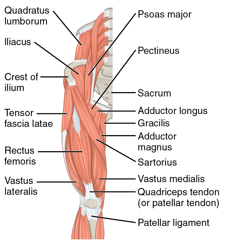
The adductor muscles form the fleshy mass on the medial side of the thigh.
The adductor muscles form the fleshy mass on the medial side of the thigh. Похожие запросы для upper thigh anatomy ct. Muscle and tendon characteristics classic human anatomy in motion: One further muscle of the anterior knee is the small articularis genus muscle, it is occasionally is blended with vastus intermedius. These images are arranged in radiographic view. Nestled into the top part of the pelvic bone, the iliacus muscle extends all the way from your lowest rib to your thigh bone. Thigh the thigh bears much of the load of the body's weight when a person is upright. It transmits the great saphenous vein, and other, smaller vessels, and is termed the fossa ovalis. Legs give us the freedom to run, walk, jump, climb, and negotiate the world around us. Human anatomy diagram 12 photos of the human anatomy diagram human anatomy body parts test, human anatomy diagram kidney location, human anatomy internal organs diagram+female, human anatomy list parts, human anatomy throat diagram, human muscles, human anatomy body parts test, human anatomy diagram kidney location. There are five muscles in the anterior thigh compartment: 1 article features images from this case. These are the gluteus maximus, gluteus medius, gluteus minimus, and tensor fasciae latae.
They work closely with your quadriceps muscles at the front of your thigh, your gluteal muscles, and your calf muscles to ensure proper movement of your leg and hip. Anatomy of the thigh and leg the thigh is best described in terms of compartmental anatomy, and is composed of anterior, posterior, and medial (adductor) compartments. In clinical anatomy the thigh muscles are divided into three groups: When you bend, run, walk, sit, or dance, the iliacus muscle works together with the other muscles in your hip joint to keep you moving smoothly and without pain. There are five muscles in the anterior thigh compartment:
Leg Pain Symptoms Treatments Causes from images.emedicinehealth.com Small and deep muscles which mainly externally rotate the thigh at the hip joint and stabilize the pelvis. It transmits the great saphenous vein, and other, smaller vessels, and is termed the fossa ovalis. The dorsal (from latin dorsum 'back') surface of an organism refers to the back, or upper side, of an organism. One further muscle of the anterior knee is the small articularis genus muscle, it is occasionally is blended with vastus intermedius. Related posts of muscle anatomy of upper thigh. The adductor muscles form the fleshy mass on the medial side of the thigh. In clinical anatomy the thigh muscles are divided into three groups: Muscle and tendon characteristics classic human anatomy in motion:
Case contributed by dr roberto schubert.
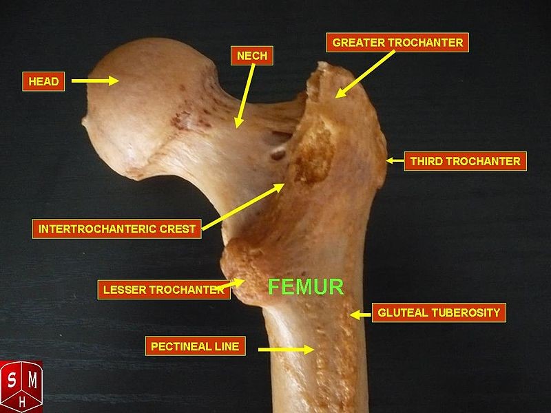
The thigh muscles don't just move your legs. It contains many muscles and nerves but only has one bone, the femur, which is the longest and strongest bone. Muscles of the leg and foot classic human anatomy in motion: The dorsal (from latin dorsum 'back') surface of an organism refers to the back, or upper side, of an organism. Human anatomy diagram 12 photos of the human anatomy diagram human anatomy body parts test, human anatomy diagram kidney location, human anatomy internal organs diagram+female, human anatomy list parts, human anatomy throat diagram, human muscles, human anatomy body parts test, human anatomy diagram kidney location. Is there an easy way to learn their a. Like the forearm, the upper leg, or thigh, has a dense arrangement of many muscles. These are the gluteus maximus, gluteus medius, gluteus minimus, and tensor fasciae latae. Large and superficial muscles which mainly abduct and extend the thigh at the hip joint. 9 public playlist includes this case. One of the most important tendons in terms of mobility of the leg is the achilles tendon. The muscles of the thigh and gluteal region are a group of complex muscles that help move and stabilize the lower limb. Upper leg anatomy and function the upper leg is often called the thigh.
Exposure variables) in a population at a given point in time. Is there an easy way to learn their a. Lewis (1918) gray's anatomy 20th ed (in public domain at yahoo or bartleby) images: This common rash often appears in the spring and fall, with symptoms including small, scaly patches on the thighs, neck, upper arms, back, or chest. Diagnosis not applicable diagnosis not applicable.
Issues Around The Hip From Tendonitis To Bursitis Beacon Orthopaedics Sports Medicine from www.beaconortho.com Thigh anatomy is fairly complex, since we are talking about two of the main joints in the human body: When you bend, run, walk, sit, or dance, the iliacus muscle works together with the other muscles in your hip joint to keep you moving smoothly and without pain. They have a lot to do with how your hips move. Exposure variables) in a population at a given point in time. Ebraheim's educational animated video describes muscle anatomy of the thigh. If talking about the skull, the dorsal side is the top. There are five muscles in the anterior thigh compartment: Check out the diagram of leg muscle anatomy in the first diagram below.
Anatomy of the thigh and leg the thigh is best described in terms of compartmental anatomy, and is composed of anterior, posterior, and medial (adductor) compartments.
On the anterior side, the most prominent of the muscles are the sartorius muscle and the four muscles that make up quadriceps muscle group (the quads.) These images are arranged in radiographic view. Related posts of muscle anatomy of upper thigh. The muscles of the thigh and gluteal region are a group of complex muscles that help move and stabilize the lower limb. It transmits the great saphenous vein, and other, smaller vessels, and is termed the fossa ovalis. Case contributed by dr roberto schubert. Thigh the thigh bears much of the load of the body's weight when a person is upright. Sartorius, and the four quadriceps muscles; These two terms, used in anatomy and embryology, describe something at the back (dorsal) or front/belly (ventral) of an organism. The muscles located within the posterior compartment of the thigh are the biceps femoris, semitendinosus and semimembranosus. In clinical anatomy the thigh muscles are divided into three groups: The four muscles all extend the lower leg. Iliopsoas muscle, a hip flexor muscle that attaches to the upper thigh bone.